

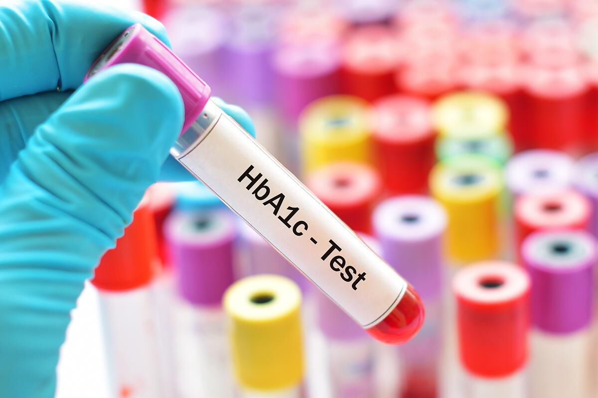

Have you ever wondered how doctors can identify inherited blood disorders by examining a small blood sample? Modern laboratory techniques make it possible to detect different types of hemoglobin and identify abnormalities that may affect the body's ability to transport oxygen.
One important investigation used for this purpose is the Hemoglobin Electrophoresis Test in Dubai. It is a specialized blood test that separates different hemoglobin proteins according to their electrical properties. By examining the separated proteins, laboratory specialists can identify abnormal hemoglobin variants and measure their percentages.
Healthcare professionals commonly recommend this test when investigating unexplained anemia, suspected sickle cell disease, thalassemia, or a family history of inherited blood disorders.
But how does the process actually work? What happens to your blood sample inside the laboratory, and how do specialists interpret the final results?
This article explains the science, testing procedure, laboratory techniques, result interpretation, and medical importance of hemoglobin electrophoresis.
The Basic Principle: How Does Hemoglobin Electrophoresis Work?
Hemoglobin electrophoresis works through a scientific process called electrophoresis, which uses an electrical field to separate proteins according to their electrical characteristics.
Hemoglobin is an iron-containing protein found inside red blood cells. Its primary function is to transport oxygen from the lungs to different tissues and organs.
Normal adult blood mainly contains hemoglobin A. However, inherited genetic changes can produce different hemoglobin structures, including hemoglobin S, hemoglobin C, and hemoglobin E.
These hemoglobin variants have different electrical properties. When exposed to an electrical field under controlled laboratory conditions, they migrate through a supporting medium at different rates.
This separation creates identifiable patterns that allow laboratory professionals to distinguish between normal and abnormal hemoglobin types.
In simple terms, the test does not directly examine how much oxygen your blood carries. Instead, it examines the types of hemoglobin proteins present in your blood.
From Blood Sample to Final Report: The Complete Testing Process
Although the laboratory science behind hemoglobin analysis is specialized, the procedure is straightforward for patients.
Understanding each stage helps explain how a simple blood collection becomes a detailed diagnostic report.
Step 1: Collecting the Blood Sample
The process begins when a healthcare professional collects a small blood sample, usually from a vein in your arm.
The skin is cleaned with an antiseptic solution before a sterile needle is inserted. The blood is collected in an appropriate laboratory tube.
For newborns, blood may be collected through a heel-prick procedure as part of newborn screening.
The sample is then labeled and transported to the laboratory for further processing.
Step 2: Preparing the Hemoglobin Sample
Once the blood reaches the laboratory, specialists prepare it for hemoglobin analysis.
Red blood cells are processed to release their hemoglobin content. This preparation provides the material required to identify and separate the different hemoglobin fractions.
Proper sample handling is essential because laboratory quality and sample integrity can influence the accuracy of the results.
Step 3: Applying an Electrical Field
The prepared hemoglobin sample is placed on an appropriate supporting medium, depending on the electrophoresis technique being used.
An electrical current is applied across the medium.
Because different hemoglobin proteins have different electrical charges and molecular characteristics, they migrate at different rates under the selected testing conditions.
This process separates the hemoglobin fractions into identifiable patterns.
Step 4: Identifying Different Hemoglobin Types
After separation, laboratory specialists examine the resulting pattern.
They look for different hemoglobin fractions, including:
-
Hemoglobin A (HbA)
-
Hemoglobin A2 (HbA2)
-
Hemoglobin F (HbF)
-
Hemoglobin S (HbS)
-
Hemoglobin C (HbC)
The presence or absence of particular fractions provides important information about normal and abnormal hemoglobin production.
Step 5: Measuring Hemoglobin Percentages
Modern laboratory methods can quantify the amount of each identified hemoglobin type.
The results are generally presented as percentages of total hemoglobin.
For example, an adult with a typical hemoglobin pattern will usually have predominantly HbA, smaller amounts of HbA2, and very little HbF.
Abnormal percentages may suggest an inherited hemoglobin condition that requires additional evaluation.
Step 6: Preparing the Laboratory Report
The final report includes the identified hemoglobin types and their measured percentages.
A laboratory specialist verifies the findings, and the report is provided to the requesting healthcare professional.
Your doctor interprets the results alongside your complete blood count, medical history, symptoms, and family history.
Which Laboratory Techniques Are Used to Analyze Hemoglobin?
Hemoglobin electrophoresis is not the only technique used to identify abnormal hemoglobin proteins. Laboratories may use several analytical methods depending on the clinical requirements and available equipment.
Traditional Hemoglobin Electrophoresis
Traditional electrophoresis separates hemoglobin fractions using an electrical field.
Different variants migrate through a supporting medium according to their electrical properties.
The resulting pattern helps specialists identify different hemoglobin types and investigate inherited blood disorders.
High-Performance Liquid Chromatography (HPLC)
HPLC is a widely used laboratory technique for separating and measuring hemoglobin fractions.
It uses differences in how hemoglobin components interact with a specialized column.
The technique produces a chromatogram showing different hemoglobin fractions and their measured percentages.
It is particularly useful for investigating beta-thalassemia, sickle cell conditions, and other hemoglobin variants.
Capillary Electrophoresis
Capillary electrophoresis separates hemoglobin proteins inside narrow capillary tubes using an electrical field.
This technique can provide detailed separation and measurement of different hemoglobin fractions.
It is used in many modern laboratories because it offers useful analytical information and can help distinguish certain variants.
Although these methods use different scientific principles, they share the same general purpose: identifying and measuring the different types of hemoglobin present in a blood sample.
What Happens When Different Hemoglobin Types Are Separated?
The separation process helps healthcare professionals understand whether hemoglobin production follows a typical adult pattern or suggests an inherited condition.
Here is what the main hemoglobin fractions can reveal.
Hemoglobin A (HbA): This is the primary adult hemoglobin and generally accounts for approximately 95% to 98% of total adult hemoglobin.
Hemoglobin A2 (HbA2): This minor adult component usually represents approximately 2% to 3.5%. Increased levels may suggest beta-thalassemia trait.
Hemoglobin F (HbF): Also known as fetal hemoglobin, HbF is naturally abundant before birth. Higher levels in adults may occur in certain inherited conditions.
Hemoglobin S (HbS): This abnormal variant is associated with sickle cell trait and sickle cell disease.
Hemoglobin C (HbC): This inherited variant may occur as a carrier condition or in combination with other abnormal hemoglobin types.
Other variants, such as hemoglobin E, may also be identified depending on the analytical method.
How Do Doctors Interpret Hemoglobin Electrophoresis Results?
After the laboratory completes the analysis, the doctor examines the hemoglobin percentages and compares them with the expected reference intervals.
The following table provides general examples of adult hemoglobin patterns.
| Hemoglobin Type | Typical Adult Level |
|---|---|
| HbA | 95–98% |
| HbA2 | 2–3.5% |
| HbF | Usually below 1–2% |
| HbS | Absent |
| HbC | Absent |
These are approximate educational ranges. Actual laboratory reference intervals may differ according to age and analytical technique.
Understanding Abnormal Hemoglobin Patterns
Certain findings may indicate inherited blood disorders.
Increased HbA2: May suggest beta-thalassemia trait, especially when accompanied by a low mean corpuscular volume (MCV).
Presence of HbS: May indicate sickle cell trait or sickle cell disease. The overall distribution of hemoglobin fractions helps distinguish between different patterns.
Increased HbF: May occur in thalassemia, hereditary persistence of fetal hemoglobin, and certain other conditions.
Presence of HbC: May suggest hemoglobin C trait or hemoglobin C disease, depending on the complete laboratory pattern.
An abnormal result does not automatically confirm a disease. Additional testing may be necessary to establish the diagnosis.
Why Is Hemoglobin Electrophoresis Important for Detecting Blood Disorders?
The test plays an important role in investigating inherited conditions that affect red blood cell function.
Identifying Sickle Cell Disease
Sickle cell disease develops when a person inherits specific combinations of abnormal hemoglobin genes.
Hemoglobin S can cause red blood cells to become rigid and sickle-shaped, potentially leading to anemia, painful episodes, and other complications.
Hemoglobin analysis helps identify the different patterns associated with sickle cell conditions.
Investigating Thalassemia
Thalassemia affects the production of alpha or beta globin chains.
Hemoglobin electrophoresis is particularly useful in identifying beta-thalassemia patterns, including increased HbA2.
Alpha-thalassemia may require additional molecular testing because some carrier forms can have normal adult electrophoresis findings.
Supporting Genetic and Pregnancy Screening
The test can identify individuals who carry inherited hemoglobin variants.
When both prospective parents carry certain relevant genetic changes, their children may have an increased risk of developing an inherited blood disorder.
Screening and genetic counseling help families understand these risks and available testing options.
Factors That May Affect the Accuracy of Results:
Although hemoglobin electrophoresis is a valuable laboratory investigation, several factors can influence result interpretation.
Recent blood transfusions: Donor red blood cells may introduce different hemoglobin types and alter measured percentages.
Age: Newborns naturally have higher fetal hemoglobin levels than adults.
Iron deficiency: Iron deficiency can complicate the interpretation of HbA2 levels in some patients.
Similar hemoglobin migration patterns: Certain variants may appear similar using particular electrophoresis methods and require additional analytical techniques.
For these reasons, healthcare professionals may recommend HPLC, capillary electrophoresis, or genetic testing when findings are unclear.
Does the Patient Need Special Preparation Before Testing?
In most cases, hemoglobin electrophoresis requires very little preparation.
Patients generally do not need to fast before providing a blood sample unless other tests are being performed simultaneously.
However, you should inform your healthcare provider about recent blood transfusions, previous abnormal blood results, family history of inherited blood disorders, and relevant medical conditions.
The blood collection procedure is generally safe. Temporary discomfort, mild bruising, or slight bleeding may occur at the collection site.
Frequently Asked Questions:
1. How long does hemoglobin electrophoresis take?
Blood collection usually takes only a few minutes. Laboratory processing and reporting may take several days, depending on the testing method and facility.
2. Does hemoglobin electrophoresis detect anemia?
The test does not measure anemia in the same way as a complete blood count. However, it can identify inherited hemoglobin abnormalities that may contribute to anemia.
3. Can the test identify thalassemia?
Yes. It can help identify beta-thalassemia patterns. Additional investigations may be required to diagnose alpha-thalassemia.
4. Is hemoglobin electrophoresis painful?
No significant pain is usually involved. Most patients experience only brief discomfort during routine blood collection.
5. Can a healthy person have abnormal hemoglobin?
Yes. Some people carry inherited hemoglobin variants without experiencing noticeable symptoms. Their results may still be important for family planning.
6. What happens if the results are abnormal?
Your doctor may recommend additional blood investigations, genetic testing, or consultation with a hematologist to determine the significance of the findings.
Final Thoughts: Understanding the Technology Behind Hemoglobin Testing
The hemoglobin electrophoresis Lab tests works by separating different hemoglobin proteins according to their electrical properties, allowing laboratory specialists to identify and measure normal and abnormal hemoglobin fractions.
From collecting a small blood sample to producing a detailed laboratory report, each stage contributes to identifying inherited blood disorders and investigating unexplained anemia.
The test is particularly valuable for detecting hemoglobin patterns associated with sickle cell disease and beta-thalassemia. It also supports newborn screening, genetic counseling, and pregnancy-related investigations.
Although the procedure is simple, accurate interpretation requires consideration of age, recent blood transfusions, iron status, and other laboratory findings.
If your healthcare provider recommends this investigation, discuss the results with a qualified medical professional. Understanding how the test works can help you make informed decisions about your blood health and any additional care you may need.